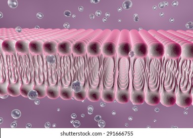
The size of the virus-containing EVs varies widely among different viruses, as does the number of virus capsids enclosed in each vesicle, most likely reflecting different mechanisms of biogenesis. This phenomenon was recognized first among members of the Picornaviridae, including hepatitis A virus (HAV, genus Hepatovirus) ( Feng et al., 2013), poliovirus and coxsackievirus B (genus Enterovirus) ( Bird et al., 2014 Chen et al., 2015 Jackson et al., 2005 Robinson et al., 2014), but it has been demonstrated also for hepatitis E virus ( Hepeviridae), rotaviruses ( Reoviridae), and noroviruses ( Caliciviridae) ( Nagashima et al., 2017 Santiana et al., 2018). However, many viruses that have previously been considered to be ‘non-enveloped’ are now known to be released non-lytically from infected cells in a ‘quasi-enveloped’ form, enclosed in small extracellular vesicles (EVs) devoid of virus-encoded surface proteins. The presence or absence of an external lipid envelope has featured strongly in the systematic classification of animal viruses for decades. Studying how these particles are able to infect human cells while hiding behind membranes borrowed from the host may help us target these viruses better. Several viruses, such as the one that causes polio, also have quasi-enveloped forms. This caused the quasi-enveloped viruses to fall apart and release their RNA into the cell more slowly than the naked particles. However, the vesicles that carried quasi-enveloped virus travelled further into the cell and eventually delivered their contents to a specialized compartment, the lysosome, where the virus membrane was degraded. Inside these vesicles, the naked virus particles soon fell apart, and their RNA was released directly into the interior of the cell. First, the external membrane of the cell folded around the particles, creating a vesicle that trapped the viruses and brought them within the cell. The experiments showed that both types of virus particle actually use similar routes. used a microscopy approach to observe Hepatitis A particles infecting human liver cells. To address this question, Rivera-Serrano et al. It was not clear how these two different types of virus particle are both able to enter cells despite their surface being so different. This membrane protects the protein shell from human immune responses, enabling quasi-enveloped virus particles to spread in a stealthy fashion within the liver. In a quasi-enveloped particle, the RNA and protein shell are completely enclosed within a membrane that is released from the host cell. Naked virus particles are shed in the feces of infected individuals and are very stable, allowing the virus to spread in the environment to find new hosts.Īt the same time, a second type of particle, known as the ‘quasi-enveloped’ virus, circulates in the blood of the infected individual. The first, known as ‘naked’ virus particles, consist of molecules of ribonucleic acid (or RNA for short) that are surrounded by a protein shell. Infected human cells produce two different types of Hepatitis A particles.

It is unable to multiply on its own so it needs to enter the cells of its host and hijack them to make new virus particles. The Hepatitis A virus is a common cause of liver disease in humans. Thus naked and quasi-enveloped virions enter via similar endocytic pathways, but uncoat in different compartments and release their genomes to the cytosol in a manner mechanistically distinct from other Picornaviridae. Neither virion requires PLA2G16, a phospholipase essential for entry of other picornaviruses. Uncoating of naked virions occurs in late endosomes, whereas eHAV undergoes ALIX-dependent trafficking to lysosomes where the quasi-envelope is enzymatically degraded and uncoating ensues coincident with breaching of endolysosomal membranes. We show both virion types enter by clathrin- and dynamin-dependent endocytosis, facilitated by integrin β 1, and traffic through early and late endosomes. eHAV lacks virus-encoded surface proteins, and how it enters cells is unknown. Quasi-enveloped HAV (eHAV) mediates stealthy cell-to-cell spread within the liver, whereas stable naked virions shed in feces are optimized for environmental transmission. Many ‘non-enveloped’ viruses, including hepatitis A virus (HAV), are released non-lytically from infected cells as infectious, quasi-enveloped virions cloaked in host membranes.


 0 kommentar(er)
